SKELETAL MUSCLES CREATE MOVEMENT
The primary function of skeletal muscle is to produce voluntary gross and fine movements to keep you alive. Large movements include walking, standing, gathering food, cooking food, turning in a chair, running, playing sports and lifting weights. Fine motor skills or smaller movements include chewing, closing your eyes, blinking, typing, writing and talking. Your skeletal muscles will also contract as a reflex to stimuli, like moving your hand from a very hot coffee cup or blinking your eyes when an eyelash lands on the surface of the eye.
SKELETAL MUSCLES PROTECT ORGANS
The abdominal muscles and the muscles of your lower back help to protect your vital organs. Your abdominal cavity is not protected by bones in the way that your rib cage protects your heart and lungs. Your rectus abdominus, or "six pack" muscle; your obliques, found at the sides of your torso; and your transverse abdominus, running side to side across the front of your abdominal cavity, protect your organs from the front and sides. From the lower back, your lats, quadrates lumborum and your psoas muscles, which run from the bottom area of your ribs to your pelvic bones, protect your organs from the back of your abdominal cavity.
Cardiac Muscle Pumps Blood
The contraction of the heart muscle is involuntary and primarily controlled by your heart's own electrical system, with and without influence from factors in the blood. Your heart is responsible for receiving blood back from your muscles, pumping it into your lungs, receiving the blood from the lungs then pumping it out into your arteries to supply your entire body. When your heart muscle does not receive enough blood due to dead heart tissue or lack of sufficient oxygen, you will have a heart attack.
Smooth Muscle Aids Digestion
The smooth muscles in your stomach and intestines work to process the food you eat. The involuntary contractions in your stomach and intestines aid in digestion and in moving the food along your digestive tract, ultimately directing indigestible substances to your rectum.
Smooth Muscle Ensures Blood Flow
There are smooth muscles in the walls of your blood vessels. When your heart contracts, your arteries expand to accept the blood. The smooth muscles in your arteries contract to push the blood throughout the blood vessel systems in your body, ultimately pushing the blood from your arterioles into your capillaries to return back to the heart. When plaque builds up on the walls of your arteries, your arteries harden and the muscles in your arteries will not contract properly.
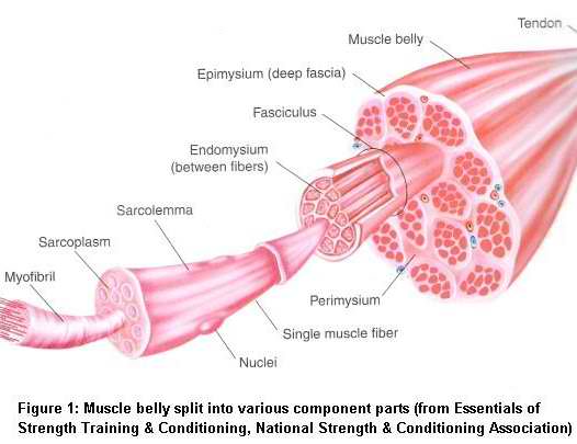
Parts Of The Muscle
Limb muscles (such as the biceps brachii in
the upper arm) have two attachments to bone.
The proximal or origin is the attachment
closest to the trunk. The distal or insertion
is the attachment furthest from the trunk.
Trunk muscles (such as the rectus abdominus
in the stomach) also have two attachments -
superior (closer to the head) and inferior
(further from the head).
A closer look at muscle anatomy shows that
each muscle belly is made up of muscle cells
or fibres. Muscle fibres are grouped into
bundles (of up to 150 fibres) called
fasciculi. Each fasiculus or bundle is
surrounded by connective tissue called
perimysium. Fibres within each bundle are
surrounded by more connective tissue called
endomysium.
Each individual fibre consists of a membrane
(sarcolemma) and can be further broken down
into hundreds or even thousands of myofibrils.
Myofibrils are surrounded by sarcoplasm and
together they make up the contractile
components of a muscle. Sarcoplasm contains glycogen, fat particles, enzymes and the mitochondria. The myofibrils it encases consist of two types of protien filaments or myofilaments. They are actin and myosin.
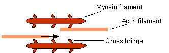
Myosin and actin filaments run in parallel to each other along the length of the muscle fibre. Myosin has tiny globular heads protruding from it at regular intervals. These are called cross bridges and play a pivotal role in muscle action. For more details, see the article on sliding filament theory.
Each myofibril is organized into sections along its length. Each section is called a sarcomere and they are repeated right along the length of a muscle fibre. It's similar to how a meter ruler is split into centimeters and millimeters. Just as the millimeter is the smallest function of a ruler, the sarcomere is the smallest contractile portion of a muscle fibre.
The sarcomere is often divided up into different zones to show how it behaves during muscle action. The Z-line seperates each sarcomere. The H-zone is the center of the sarcomere and the M-line is where adjacent myosin filaments anchor on to each other. On the diagram above the darker A-bands are where myosin filaments align and the lighter I-bands are where actin filaments align. When muscle contracts the H-zone and I-band both decrease as the z-lines are pulled towards each other. When actin filaments (the light bands in the diagram above) slide over myosin filaments (the dark bands) the H-zone and I-band decrease.
What causes actin filaments to move?
Myosin filaments contain tiny globular heads, called cross bridges at regular intervals. These cross bridges attach to the actin filaments pulling on them to create movement. The Sarcoplasmic Reticulum
Surrounding each myofibril (remember a myofibril is the portion of the muscle fibre that houses actin and myosin) is a system of tubules called the sarcoplasmic reticulum. The sarcoplasmic reticulum stores calcium and it is the regluation of calcium release that causes muscular contraction.
During rest most of the calcium resides in the sarcoplasmic reticulum and and very few myosin cross bridges are attached to actin filaments - nor can they flex.
When the brain sends a nerve impulse (called an action potential)
How does that cause a muscle fibre to contract?
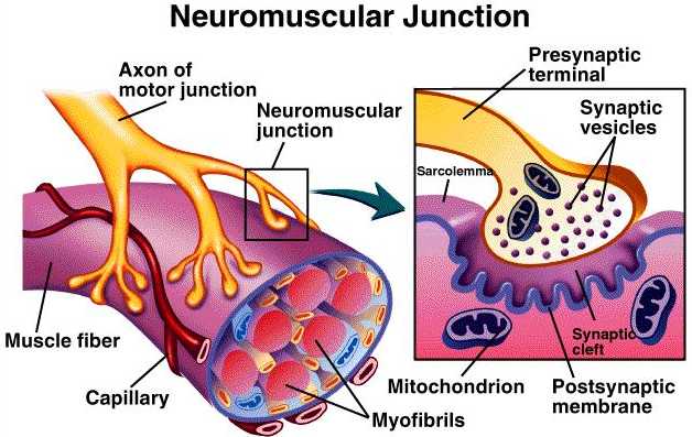
The action potential arrives at the nerve terminal and causes the release of a chemical called acetylcholine. Acetylcholine travels across the neuromusclualr junction and stimulates the sarcoplasmic reticulum to release its stored calcium ions throughout the muscle.
Excitation-Contraction Coupling
As calcium is relased it binds with a protein called troponin that is situated along the actin filaments. Sliding filament theory states that this binding causes a shift to occur in another chemical called tropomyosin. Because these chemicals have a high affinity for calcium ions they cause the myosin cross bridges to attach to actin and flex rapidly.
For contraction to contiune the myosin cross bridges must detach, "recock" and reattach. Significant muscle shortening depends on the continuous sequence of the following events:
Calcium released by sarcoplasmic reticulum binds with troponin
Myosin cross bridge couples with actin filament
Cross bridge flexes and moves actin a small amount
Cross bridge detaches and re-cocks
Process is repeated
The Acetylcholine Receptor and the Neuromuscular Junction
One of the best-studied examples of chemical transmission is the neuromuscular junction, the synapse between a motor neuron and a skeletal muscle fiber. Synaptic transmission is mediated by the neurotransmitter acetylcholine, which is released into the synaptic cleft by the presynaptic neuron. In the synaptic cleft, acetylcholine either binds to its receptor on the postsynaptic cell or is rapidly degraded by the enzyme acetylcholinesterase. This ensures that there is only a brief response of the postsynaptic cell. The acetylcholine receptoris a relatively nonspecific ligand-gated ion channel that is open to the flow of ions only when it binds to acetylcholine. Upon binding to acetylcholine, the muscle fiber is stimulated. There is an influx of sodium ions into the postsynaptic muscle fiber and the membrane becomes depolarized, leading to an action potential, and ultimately, muscle contraction. The nerve gas sarin is a potent neurotoxin and inhibitor of acetylcholineseterase, which affects an accumulation of acetylcholine at the neuromuscular junction. The high levels of acetylcholine in the synaptic cleft lead to increased depolarization and contraction of the muscle fibers; however, without the ability to recover from stimulation, the muscle fiber is inactivated, thus leading to paralysis. CLICK HERE FOR ANIMATION OF ACTION POTENTIAL CLICK HERE FOR ANATOMY OF MUSCLES <<RETURN TO TOPICS>>