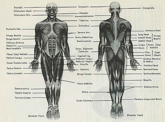
ANATOMY OF MUSCLES anterior and posterior view of superficial body musculature
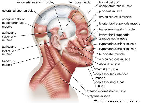
click image to enlarge Muscles of Facial Expression
The muscles of facial expression originate on the surface of the skull. At their insertions, the fibers of the epimysium are woven into those of the superficial fascia and the dermis of the skin: When they contract, the skin moves. These muscles are innervated by the facial nerve.
The largest group of facial muscles is associated with the mouth. The orbicularis oris muscle constricts the opening, and other muscles move the lips or the corners of the mouth. The buccinator muscle has two functions related to eating (in addition to its importance to musicians). During chewing, it cooperates with the masticatory muscles by moving food back across the teeth from the space inside the cheeks. In infants, the buccinator provides suction for suckling at the breast.
Smaller groups of muscles control movements of the eyebrows and eyelids, the scalp, the nose, and the external ear. The epicranius, or scalp, contains two muscles, the frontalis and the occipitalis muscles. These muscles are separated by the galea aponeurotica, a collagenous sheet. The platysma covers the ventral surface of the neck, extending from the base of the neck to the periosteum of the mandible and the fascia at the corner of the mouth.
xtrinsic Eye Muscles
Six extrinsic eye muscles, or oculomotor muscles, originating on the surface of the orbit control the position of each eye. These are the inferior rectus, medial rectus, superior rectus, lateral rectus, inferior oblique, and superior oblique muscles. The extrinsic eye muscles are innervated by the third (oculomotor), fourth (trochlear), and sixth (abducens) cranial nerves.
Muscles of Mastication
The muscles of mastication move the mandible at the temporomandibular joint. The large masseter muscle is the strongest jaw muscle. The temporalis muscle assists in elevation of the mandible. The pterygoid muscles, used in various combinations, can elevate, depress, or protract the mandible or slide it from side to side, a movement called lateral excursion. These movements are important in making efficient use of your teeth while you chew foods of various consistencies. The muscles of mastication are innervated by the fifth cranial nerve, the trigeminal nerve.
Muscles of the Tongue
The muscles of the tongue have names ending in glossus, the Greek word for "tongue." Once you can recall the structures referred to by palato-, stylo-, genio-, and hyo-, you will follow this group. The palatoglossus muscle originates at the palate, the styloglossus muscle at the styloid process of the temporal bone, the genioglossus muscle at the chin, and the hyoglossus muscle at the hyoid bone. These muscles, used in various combinations, move the tongue in the delicate and complex patterns necessary for speech, and they manipulate food within the mouth in preparation for swallowing. Most are innervated by the hypoglossal nerve , a cranial nerve whose name indicates its function and its location. (Table 11-5)
Muscles of the Pharynx
The muscles of the pharynx are responsible for initiating the swallowing process. The pharyngeal constrictors (superior, middle, and inferior) move materials into the esophagus. The laryngeal elevators elevate the larynx. The palatal muscles, the tensor veli palatini and the levator veli palatini, raise the soft palate and adjacent portions of the pharyngeal wall and also pull open the entrance to the auditory tube. As a result, swallowing repeatedly can open the entrance to the auditory tube to help you adjust to pressure changes when you fly or dive.
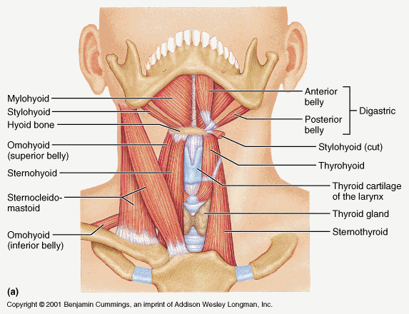
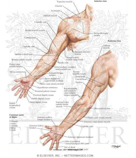
Anterior Muscles of the Neck
The anterior muscles of the neck include (1) muscles that control the position of the larynx, (2) muscles that depress the mandible and tense the floor of the mouth, and (3) muscles that provide a stable foundation for muscles of the tongue and pharynx (Figure 11-10 and Table 11-7). The digastric muscle has two bellies, as the name implies. One belly extends from the chin to the hyoid bone, and the other continues from the hyoid bone to the mastoid portion of the temporal bone. Depending on which belly contracts and whether fixator muscles are stabilizing the position of the hyoid bone, the digastric muscle can open the mouth by depressing the mandible, or it can elevate the larynx by raising the hyoid bone. The digastric muscle overlies the broad, flat mylohyoid muscle, which provides a muscular floor to the mouth. The stylohyoid muscle forms a muscular connection between the hyoid bone and the styloid process of the skull. The sternocleidomastoid muscle extends from the clavicle and the sternum to the mastoid region of the skull . The omohyoid muscle attaches to the scapula, the clavicle and first rib, and the hyoid bone. The other members of this group are straplike muscles that extend between the sternum and larynx (sternothyroid) or hyoid bone (sternohyoid), between the larynx and hyoid bone (thyrohyoid), and between the hyoid bone and chin (geniohyoid). hyoid muscle deep neck and back muscle
function:
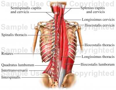
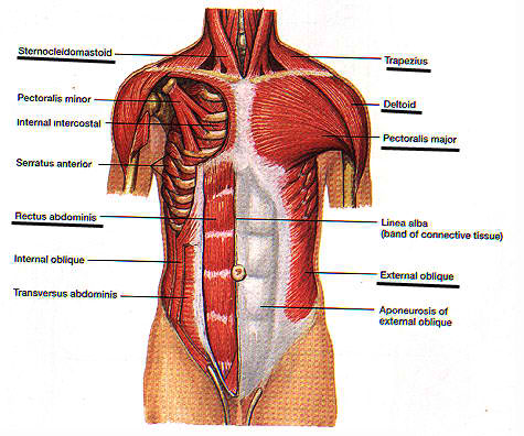
The intricate anatomy of the back provides support for the head and trunk of the body, strength in the trunk of the body, as well as a great deal of flexibility and movement. The upper back has the most structural support, with the ribs attached firmly to each level of the thoracic spine and very limited movement. The lower back (lumbar vertebrae) allows for flexibility and movement in back bending (extension) and forward bending (flexion). It does not permit twisting. arm muscles chest and abdominal muscle lower limb muscle <<RETURN TO TOPICS>>