The lymphatic system is, first of all, a part of the circulatory system, comprising a network of conduits called lymphatic vessels that carry a clear fluid called lymph unidirectionally toward the heart. The lymphatic system was first described independently by Olaus Rudbeck and Thomas Bartholin.
Secondly the lymphatic organs play an important part in the immune system, having a considerable overlap with the lymphoid system. Lymphoid tissue is found in many organs, particularly the lymph nodes, and in the lymphoid follicles associated with the digestive system such as the tonsils. The system also includes all the structures dedicated to the circulation and production of lymphocytes, which includes the spleen, thymus, bone marrow and the lymphoid tissue associated with the digestive system.
The blood does not directly come in contact with the parenchymal cells and tissues in the body, but constituents of the blood first exit the microvascular exchange blood vessels to become interstitial fluid, which comes into contact with the parenchymal cells of the body. Lymph is the fluid that is formed when interstitial fluid enters the initial lymphatic vessels of the lymphatic system. The lymph is then moved along the lymphatic vessel network by either intrinsic contractions of the lymphatic passages or by extrinsic compression of the lymphatic vessels via external tissue forces (e.g. the contractions of skeletal muscles). Eventually, the lymph vessels empty into the lymphatic ducts, which drain into one of the two subclavian veins (near the junctions of the subclavian veins with the internal jugular veins).
TONSILS
Tonsils,most commonly, the palatine tonsils that can be seen in the back of the human throat. The palatine tonsils and the nasopharyngeal tonsil are lymphoepithelial tissues located near the oropharynx and nasopharynx. These immunocompetent tissues are the immune system's first line of defense against ingested or inhaled foreign pathogens. However, the fundamental immunological roles of tonsils have yet to be understood.
Tonsillitis is a disorder in which the tonsils are inflamed (sore and swollen). The most common way to treat it is with anti-inflammatory drugs such as ibuprofen, or if bacterial in origin, antibiotics. Many sufferers treat it by having their tonsils surgically removed by a tonsillectomy. LYMPH NODES
A lymph node is a small ball or an oval-shaped organ of the immune system, distributed widely throughout the body including the armpit and stomach/gut and linked by lymphatic vessels. Lymph nodes are garrisons of B, T, and other immune cells. Lymph nodes are found all through the body, and act as filters or traps for foreign particles. They are important in the proper functioning of the immune system. They are packed tightly with the white blood cells called lymphocytes and macrophages.
Lymph nodes also have clinical significance. They become inflamed or enlarged in various conditions, which may range from trivial, such as a throat infection, to life-threatening such as cancers. In the latter, the condition of lymph nodes is so significant that it is used for cancer staging, which decides the treatment to be employed, and for determining the prognosis.
Lymph nodes can also be diagnosed by biopsy whenever they are inflamed. Certain diseases affect lymph nodes with characteristic consistency and location.
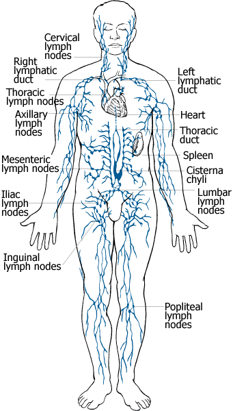
Humans have approximately 500-600 lymph nodes distributed throughout the body, with clusters found in the underarms, groin, neck, chest, and abdomen.
Lymph nodes of the head and neck
Cervical lymph nodes
Lymph nodes of the thorax
Lymph nodes of the lungs: The lymph is drained from the lung tissue through subsegmental, segmental, lobar and interlobar lymph nodes to the hilar lymph nodes, which are located around the hilum (the pedicle, which attaches the lung to the mediastinal structures, containing the pulmonary artery, the pulmonary veins, the main bronchus for each side, some vegetative nerves and the lymphatics) of each lung. The lymph flows subsequently to the mediastinal lymph nodes.
Mediastinal lymph nodes: They consist of several lymph node groups, especially along the trachea (5 groups), along the esophagus and between the lung and the diaphragm. In the mediastinal lymph nodes arises lymphatic ducts, which draines the lymph to the left subclavian vein (to the venous angle in the confluence of the subclavian and deep jugular veins).
The mediastinal lymph nodes along the esophagus are in tight connection with the abdominal lymph nodes along the esophagus and the stomach. That fact facilitates spreading of tumors cells through these lymphatics in cases of cancers of the stomach and particularly of the esophagus. Through the mediastinum, the main lymphatic drainage from the abdominal organs goes via the thoracic duct (ductus thoracicus), which drains majority of the lymph from the abdomen to the above mentioned left venous angle.
Lymph nodes of the arm
These drain the whole of the arm, and are divided into two groups, superficial and deep. The superficial nodes are supplied by lymphatics that are present throughout the arm, but are particularly rich on the palm and flexor aspects of the digits.
Superficial lymph glands of the arm:
Supratrochlear glands: Situated above the medial epicondyle of the humerus, medial to the basilic vein, they drain the C7 and C8 dermatomes.
Deltoideopectoral glands: Situated between the pectoralis major and deltoid muscles inferior to the clavicle.
Deep lymph glands of the arm: These comprise the axillary glands, which are 20-30 individual glands and can be subdivided into:
Lateral glands
Anterior or pectoral glands
Posterior or subscapular glands
Central or intermediate glands
Medial or subclavicular glands
SPLEEN
The spleen is an organ found in virtually all vertebrate animals with regenerative capabilities and has important roles in regard to red blood cells (also referred to as erythrocytes) and the immune system. In humans, it is located in the left upper quadrant of the abdomen. It removes old red blood cells and holds a reserve of blood in case of hemorrhagic shock while also recycling iron. As a part of the mononuclear phagocyte system, it metabolizes hemoglobin removed from senescent erythrocytes. The globin portion of hemoglobin is degraded to its constitutive amino acids, and the heme portion is metabolized to bilirubin, which is subsequently shuttled to the liver for removal. It synthesizes antibodies in its white pulp and removes antibody-coated bacteria along with antibody-coated blood cells by way of blood and lymph node circulation. The spleen is brownish. Recently, it has been found to contain in its reserve half of the body's monocytes within the red pulp. These monocytes, upon moving to injured tissue (such as the heart), turn into dendritic cells and macrophages while promoting tissue healing. It is one of the centers of activity of the reticuloendothelial system and can be considered analogous to a large lymph node, as its absence leads to a predisposition toward certain infections.
Other functions of the spleen are less prominent, especially in the healthy adult:
Production of opsonins, properdin, and tuftsin.
Creation of red blood cells. While the bone marrow is the primary site of hematopoiesis in the adult, the spleen has important hematopoietic functions up until the fifth month of gestation. After birth, erythropoietic functions cease, except in some hematologic disorders. As a major lymphoid organ and a central player in the reticuloendothelial system, the spleen retains the ability to produce lymphocytes and, as such, remains an hematopoietic organ.
Storage of red blood cells, lymphocytes and other formed elements. In horses, roughly 30% of the red blood cells are stored there. The red blood cells can be released when needed. In humans, up to a cup (236.5ml) of red blood cells can be held in the spleen and released in cases of hypovolemia. It can store platelets in case of an emergency. Up to a quarter of lymphocytes can be stored in the spleen at any one time.
In mice, the spleen stores half the body's monocytes so that upon injury they can migrate to the injured tissue and transform into dendritic cells and macrophages and so assist wound healing.
MACROPHAGES
Macrophages are cells produced by the differentiation of monocytes in tissues. Human macrophages are about 21 micrometres (0.00083 in) in diameter. Monocytes and macrophages are phagocytes. Macrophages function in both non-specific defense (innate immunity) as well as help initiate specific defense mechanisms (adaptive immunity) of vertebrate animals. Their role is to phagocytose (engulf and then digest) cellular debris and pathogens, either as stationary or as mobile cells. They also stimulate lymphocytes and other immune cells to respond to pathogens. They are specialized phagocytic cells that attack foreign substances, infectious microbes and cancer cells through destruction and ingestion. Macrophages can be identified by specific expression of a number of proteins including CD14, CD11b, F4/80 (mice)/EMR1 (human), lysozyme M, MAC-1/MAC-3 and CD68 by flow cytometry or immunohistochemical staining. They move by action of amoeboid movement.
Phagocytosis
One important role of the macrophage is the removal of necrotic cellular debris in the lungs. Removing dead cell material is important in chronic inflammation, as the early stages of inflammation are dominated by neutrophil granulocytes, which are ingested by macrophages if they come of age (see CD-31 for a description of this process.)
The removal of necrotic tissue is, to a greater extent, handled by fixed macrophages, which will stay at strategic locations such as the lungs, liver, neural tissue, bone, spleen and connective tissue, ingesting foreign materials such as pathogens and recruiting additional macrophages if needed.
When a macrophage ingests a pathogen, the pathogen becomes trapped in a phagosome, which then fuses with a lysosome. Within the phagolysosome, enzymes and toxic peroxides digest the pathogen. However, some bacteria, such as Mycobacterium tuberculosis, have become resistant to these methods of digestion. Macrophages can digest more than 100 bacteria before they finally die due to their own digestive compounds. ANTIBODIES
An antibody, also known as an immunoglobulin, is a large Y-shaped protein produced by B-cells that is used by the immune system to identify and neutralize foreign objects such as bacteria and viruses. The antibody recognizes a unique part of the foreign target, called an antigen.Each tip of the "Y" of an antibody contains a paratope (a structure analogous to a lock) that is specific for one particular epitope (similarly analogous to a key) on an antigen, allowing these two structures to bind together with precision. Using this binding mechanism, an antibody can tag a microbe or an infected cell for attack by other parts of the immune system, or can neutralize its target directly (for example, by blocking a part of a microbe that is essential for its invasion and survival). The production of antibodies is the main function of the humoral immune system.
Antibodies are produced by type of white blood cell called a plasma cell. Antibodies can occur in two physical forms, a soluble form that is secreted from the cell, and a membrane-bound form that is attached to the surface of a B cell and is referred to as the B cell receptor (BCR). The BCR is only found on the surface of B cells and facilitates the activation of these cells and their subsequent differentiation into either antibody factories called plasma cells, or memory B cells that will survive in the body and remember that same antigen so the B cells can respond faster upon future exposure. In most cases, interaction of the B cell with a T helper cell is necessary to produce full activation of the B cell and, therefore, antibody generation following antigen binding. Soluble antibodies are released into the blood and tissue fluids, as well as many secretions to continue to survey for invading microorganisms.
Antibodies are glycoproteins belonging to the immunoglobulin superfamily; the terms antibody and immunoglobulin are often used interchangeably. Antibodies are typically made of basic structural units—each with two large heavy chains and two small light chains. There are several different types of antibody heavy chains, and several different kinds of antibodies, which are grouped into different isotypes based on which heavy chain they possess. Five different antibody isotypes are known in mammals, which perform different roles, and help direct the appropriate immune response for each different type of foreign object they encounter.
Though the general structure of all antibodies is very similar, a small region at the tip of the protein is extremely variable, allowing millions of antibodies with slightly different tip structures, or antigen binding sites, to exist. This region is known as the hypervariable region. Each of these variants can bind to a different target, known as an antigen. This enormous diversity of antibodies allows the immune system to recognize an equally wide variety of antigens. The large and diverse population of antibodies is generated by random combinations of a set of gene segments that encode different antigen binding sites (or paratopes), followed by random mutations in this area of the antibody gene, which create further diversity. Antibody genes also re-organize in a process called class switching that changes the base of the heavy chain to another, creating a different isotype of the antibody that retains the antigen specific variable region. This allows a single antibody to be used by several different parts of the immune system.
Antibodies can come in different varieties known as isotypes or classes. In placental mammals there are five antibody isotypes known as IgA, IgD, IgE, IgG and IgM. They are each named with an "Ig" prefix that stands for immunoglobulin, another name for antibody, and differ in their biological properties, functional locations and ability to deal with different antigens, as depicted in the table.
The antibody isotype of a B cell changes during cell development and activation. Immature B cells, which have never been exposed to an antigen, are known as naïve B cells and express only the IgM isotype in a cell surface bound form. B cells begin to express both IgM and IgD when they reach maturity—the co-expression of both these immunoglobulin isotypes renders the B cell 'mature' and ready to respond to antigen. B cell activation follows engagement of the cell bound antibody molecule with an antigen, causing the cell to divide and differentiate into an antibody producing cell called a plasma cell. In this activated form, the B cell starts to produce antibody in a secreted form rather than a membrane-bound form. Some daughter cells of the activated B cells undergo isotype switching, a mechanism that causes the production of antibodies to change from IgM or IgD to the other antibody isotypes, IgE, IgA or IgG, that have defined roles in the immune system.