THE INTEGUMENTARY SYSTEM
The Integumentary system has many functions:
Protects the body's internal living tissues
and organs
Protects against invasion by infectious
organisms
Protects the body from dehydration
Protects the body against abrupt changes
in temperature
Helps dispose of waste materials
Acts as a receptor for touch, pressure,
pain, heat and cold
Stores water, fat, and vitamin D. Parts Of The Skin The Epidermis STRATUM BASALE
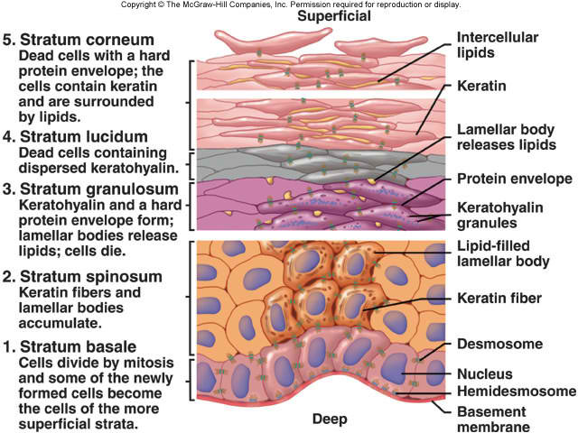
The stratum basale (basal layer, sometimes referred to as stratum germinativum) is the deepest layer of the five layers of the epidermis, which is the outer covering of skin in mammals. The stratum basale is a continuous layer of cells. It is often described as one cell thick, though it may in fact be two to three cells thick in glabrous (hairless) skin and hyperproliferative epidermis (from a skin disease).
The stratum basale is primarily made up of basal keratinocyte cells, which can be considered the stem cells of the epidermis. They divide to form the keratinocytes of the stratum spinosum, which migrate superficially.
STRATUM SPINOSUM
The stratum spinosum (or spinous layer) is a
layer of the epidermis found between the stratum granulosum and stratum basale. This layer is also referred to as the "spinous" or "prickle-cell" layer. This appearance is due to desmosomal connections of adjacent cells. Keratinization begins in the stratum spinosum.
STRATUM GRANULOSUM
The stratum granulosum (or granular layer) is a thin layer of cells in the epidermis.Keratinocytes migrating from the underlying stratum spinosum become known as granular cells in this layer. These cells contain keratohyalin granules, protein structures that promote hydration and crosslinking of keratin.
At the transition between this layer and the stratum corneum, cells secrete lamellar bodies (containing lipids and proteins) into the extracellular space. This results in the formation of the hydrophobic lipid envelope responsible for the skin's barrier properties
STRATUM LUCIDUM
The stratum lucidum (Latin for "clear layer") is a thin, clear layer of dead skin cells in the epidermis named for its translucent appearance under a microscope. It is found only in areas of thick skin, most noticeably on the palms of the hands and the soles of the feet.
Located between the stratum granulosum and stratum corneum layers, it is composed of three to five layers of dead, flattened keratinocytes. The keratinocytes of the stratum lucidum do not feature distinct boundaries and are filled with eleidin, an intermediate form of keratin.
The thickness of the lucidum is controlled by the rate of mitosis of the epidermal cells. In addition, melanocytes determine the darkness of the stratum lucidum. The cells of the stratum lucidum are flattened and contain an oily substance that is the result of exocytosis of lamellar bodies accumulated while the keratinocytes are moving through the stratum spinosum and stratum granulosum.
STRATUM CORNEUM
The stratum corneum (Latin for horned layer) is the outermost layer of the epidermis, consisting of dead cells (corneocytes) that lack nuclei and organelles. The purpose of the stratum corneum is to form a barrier to protect underlying tissue from infection, dehydration, chemicals and mechanical stress. Desquamation, the process of cell shedding from the surface of the stratum corneum, balances proliferating keratinocytes that form in the stratum basale. These cells migrate through the epidermis towards the surface in a journey that takes approximately fourteen days.
Skin Color
Melanocytes are melanin-producing cells located in the bottom layer (the stratum basale) of the skin's epidermis, the middle layer of the eye (the uvea), the inner ear, meninges,bones, and heart. Melanin is a pigment that is responsible primarily for the color of skin.
Through a process called melanogenesis, these cells produce melanin, which is a pigment found in the skin, eyes, and hair. This melanogenesis leads to a long-lasting pigmentation, which is in contrast to the pigmentation that originates from oxidation of already-existing melanin.
There are both basal and activated levels of melanogenesis; in general, lighter-skinned people have low basal levels of melanogenesis. Exposure to UV-B radiation causes an increased melanogenesis as a response to DNA photodamage. The hair
Papilla
At the base of the follicle is a large structure that is called the papilla.
The papilla is made up mainly of connective tissue and a capillary loop. Cell division in the papilla is either rare or non-existent.
Matrix
Around the papilla is the hair matrix, a collection of epithelial cells often interspersed with the pigment-producing cells, melanocytes. Cell division in the hair matrix produces the cells that will form the major structures of the hair fiber and the inner root sheath. The hair matrix epithelium is one of the fastest growing cell populations in the human body, which is why some forms of chemotherapy that kill dividing cells or radiotherapy may lead to temporary hair loss. The papilla is usually ovoid or pear shaped with the matrix wrapped completely around it except for a short stalk-like connection to the surrounding connective tissue that provides access for the capillary.
Root sheath
The root sheath is composed of an external and internal root sheath. The external root sheath appears empty with cuboid cells when stained with H&E stain. The internal root sheath is composed of three layers, Henle's layer, Huxley's layer, and an internal cuticle that is continuous with the outermost layer of the hair fiber.
Hair fiber
The hair fiber is composed of keratin.
Bulge
The bulge is located in the outer root sheath at the insertion point of the arrector pili muscle. It houses several types of stem cells, which supply the entire hair follicle with new cells, and take part in healing the epidermis after a wound.
Other structures
Other structures associated with the hair follicle include arrector pili muscles, sebaceous glands, and apocrine sweat glands. Hair follicle receptors sense the position of the hairs.
Attached to the follicle is a tiny bundle of muscle fiber called the arrector pili. This muscle is responsible for causing the follicle lissis to become more perpendicular to the surface of the skin, and causing the follicle to protrude slightly above the surrounding skin (piloerection) and a pore encased with skin oil. This process results in goose bumps (or goose flesh).
Also attached to the follicle is a sebaceous gland, which produces the oily or waxy substance sebum. The thicker the density of the hair, the more sebaceous glands that are found. NAIL
A nail is a horn-like envelope covering the
dorsal aspect of the terminal phalanges of
fingers and toes in humans
The matrix (synonyms: matrix unguis, keratogenous membrane, nail matrix, onychostroma) is the tissue (or germinal matrix) upon which the nail rests, the part of the nail bed that extends beneath the nail root and contains nerves, lymph and blood vessels. The matrix is responsible for the production of the cells that become the nail plate. The width and thickness of the nail plate is determined by the size, length, and thickness of the matrix, while the shape of the fingertip itself determines if the nail plate is flat, arched or hooked. The matrix will continue to grow as long as it receives nutrition and remains in a healthy condition. As new nail plate cells are incubated, they emerge from the matrix round and white to push older nail plate cells forward; and in this way yet older cells become compressed, flat, and translucent, making the pink colour of the capillaries in the nail bed below visible.
The lunula (occasionally called simply "the moon") is the visible part of the matrix, the whitish crescent-shaped base of the visible nail. The lunula is largest in the thumb and often absent in the little finger.
The nail bed is the skin beneath the nail plate.Like all skin, it is composed of two types of tissues: the deeper dermis, the living tissue fixed to the bone which contains capillaries and glands, and the superficial epidermis, the layer just beneath the nail plate which moves forward with the plate. The epidermis is attached to the dermis by tiny longitudinal "grooves" known as the matrix crests or crests of nail matrix (cristae matricis unguis). During old age, the plate thins and these grooves are made evident in the structure.
The nail sinus (sinus unguis) is the deep furrow into which the nail root is inserted.[
The nail root (radix unguis) is the part of nail situated in the nail sinus, i.e. the base of the nail embedded underneath the skin. It originates from the actively growing tissue below, the matrix.
The nail plate or body of nail (corpus unguis) is the actual nail, and like hair and skin, made of translucent keratin protein made of amino acids. In the nail it forms a strong flexible material made of several layers of dead, flattened cells. The plate appears pink because of the underlying capillaries. Its (transversal) shape is determined by the form of the underlying bone. In common usage, the word nail often refers to this part only.
The free margin (margo liber) or distal edge is the anterior margin of the nail plate corresponding to the abrasive or cutting edge of the nail. The hyponychium (informally known as the "quick") is the epithelium located beneath the nail plate at the junction between the free edge and the skin of the fingertip. It forms a seal that protects the nail bed. The onychodermal band is the seal between the nail plate and the hyponychium. It is found just under the free edge, in that portion of the nail where the nail bed ends and can be recognized by its glassy, greyish colour (in fair-skinned people). It is not perceptible in some individuals while it is highly prominent on others.
The eponychium is the small band of epithelium that extends from the posterior nail wall onto the base of the nail. Often and erroneously called the "proximal fold" or "cuticle", the eponychium is the end of the proximal fold that folds back upon itself to shed an epidermal layer of skin onto the newly formed nail plate. This layer of non-living, almost invisible skin is the cuticle that "rides out" on the surface of the nail plate. Together, the eponychium and the cuticle form a protective seal. The cuticle on the nail plate is dead cells and is often removed during manicure, but the eponychium is living cells and should not be touched. The perionyx is the projecting edge of the eponychium covering the proximal strip of the lunula.
The nail wall (vallum unguis) is the cutaneous fold overlapping the sides and proximal end of the nail. The lateral margin (margo lateralis) is lying beneath the nail wall on the sides of the nail and the nail groove or fold (sulcus matricis unguis) are the cutaneous slits into which the lateral margins are embedded.
The paronychium is the border tissue around the nail and paronychia is an infection in this area. <<RETURN TO TOPICS>>